Fayl:Computed tomography of human brain (5).png
Computed_tomography_of_human_brain_(5).png (512 × 512 piksel, fayl həcmi: 78 KB, MIME növü: image/png)
| Bu fayl "Vikimedia Commons"dadır və digər layihələrdə istifadə edilə bilər. |
Faylın təsvir səhifəsinə get |
| İzahComputed tomography of human brain (5).png |
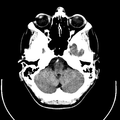
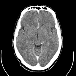
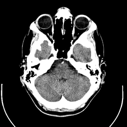
English: Computer tomography of human brain, from base of the skull to top. Taken with intravenous contrast medium.
It was taken Mars 23, 2007 on the uploader, after a 20 minute episode of homonymous hemianopsia with loss of the left visual field, but nothing strange was found. Three episodes of scotoma occurred in the following years, whereof the last one was scintillating (depiction). Otherwise, there were no further neurological symptoms.
Türkçe: Geçirdiği bir kaza neticesinde homonim hemianopsi vakası oluşan bir hastanın beyninin bilgisayarlı tomografisi. Tomografi neticesinde bir anomaliye rastlanmamıştır. |
||
| Tarix | Uploaded January 17, 2008 | ||
| Mənbə | Radiology, Uppsala University Hospital. Uploaded by Mikael Häggström. | ||
| Müəllif | Department of Radiology, Uppsala University Hospital. Uploaded by Mikael Häggström. | ||
| İcazə (Faylın təkrar istifadəsi) |
|
Mündəricat
Compound images
-
Inverted
Scrollable stack
For larger version, see Category:Computed tomography images of Mikael Häggström's brain. To move through the images, hover over the image and use scroll wheel, drag the mouse, or click the < or the > above each stack. This functionality should activate when the page is fully loaded, which may take some time.

|
The template Imagestack requires additional javascript-code. It doesn't work if javascript is switched off. |
Case with multiplanar reconstruction
-
Brain, case 1: Multiplanar, but no intravenous contrast.
Individual images
Licencing
| This file is made available under the Creative Commons CC0 1.0 Universal Public Domain Dedication. | |
| The person who associated a work with this deed has dedicated the work to the public domain by waiving all of their rights to the work worldwide under copyright law, including all related and neighboring rights, to the extent allowed by law. You can copy, modify, distribute and perform the work, even for commercial purposes, all without asking permission.
http://creativecommons.org/publicdomain/zero/1.0/deed.enCC0Creative Commons Zero, Public Domain Dedicationfalsefalse |
DICOM format
Captions
Items portrayed in this file
təsvir edir
copyright status ingilis
media type ingilis
image/png
checksum ingilis
43f3988ee01bfd2888403b7e848a1cfbc65c2167
data size ingilis
79.836 Bayt
512 piksel
512 piksel
Faylın tarixçəsi
Faylın əvvəlki versiyasını görmək üçün gün/tarix bölməsindəki tarixlərə klikləyin.
| Tarix/Vaxt | Kiçik şəkil | Ölçülər | İstifadəçi | Şərh | |
|---|---|---|---|---|---|
| indiki | 12:43, 1 fevral 2008 |  | 512 × 512 (78 KB) | Mikael Häggström | {{34 computer tomography images}} |
Fayl keçidləri
Bu faylı istifadə edən səhifə yoxdur.
Faylın qlobal istifadəsi
Bu fayl aşağıdakı vikilərdə istifadə olunur:
- fr.wikipedia.org layihəsində istifadəsi


