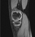
Fayl:OCD WalterReed MRI-Sagital-T1.jpeg
Naviqasiyaya keç
Axtarışa keç

Sınaq göstərişi ölçüsü: 579 × 600 piksel. Digər ölçülər: 232 × 240 piksel | 463 × 480 piksel | 945 × 979 piksel.
Faylın orijinalı (945 × 979 piksel, fayl həcmi: 353 KB, MIME növü: image/jpeg)
Faylın tarixçəsi
Faylın əvvəlki versiyasını görmək üçün gün/tarix bölməsindəki tarixlərə klikləyin.
| Tarix/Vaxt | Kiçik şəkil | Ölçülər | İstifadəçi | Şərh | |
|---|---|---|---|---|---|
| indiki | 21:10, 4 mart 2009 |  | 945 × 979 (353 KB) | FoodPuma | Added arrow (edited with Adobe Photoshop CS2) |
| 00:12, 8 fevral 2009 |  | 516 × 467 (79 KB) | FoodPuma | {{Information |Description={{en|1="Sagittal and coronal T1 and T2 images demonstrate linear low T1, high T2 signal at the articular surfaces of the lateral aspects of the medial femoral condyles bilaterally, corresponding to the radiographs, confirming th |
Fayl keçidləri
Aşağıdakı səhifə bu faylı istifadə edir:
Faylın qlobal istifadəsi
Bu fayl aşağıdakı vikilərdə istifadə olunur:
- ar.wikipedia.org layihəsində istifadəsi
- ca.wikipedia.org layihəsində istifadəsi
- en.wikipedia.org layihəsində istifadəsi
- ja.wikipedia.org layihəsində istifadəsi
- nl.wikipedia.org layihəsində istifadəsi
- pl.wikipedia.org layihəsində istifadəsi
- uk.wikipedia.org layihəsində istifadəsi
- www.wikidata.org layihəsində istifadəsi
- zh.wikipedia.org layihəsində istifadəsi

