Fayl:Respiratory system complete numbered.svg

Faylın orijinalı (SVG faylı, nominal olaraq 718 × 914 piksel, faylın ölçüsü: 507 KB)
| Bu fayl "Vikimedia Commons"dadır və digər layihələrdə istifadə edilə bilər. |
 |
Faylın təsvir səhifəsinə get |
| İzah |
[] English: Note: See the version numbered to create or enhance one translation.
|
||
| Tarix | |||
| Mənbə | Öz işi | ||
| Müəllif | LadyofHats, Jmarchn | ||
| İcazə (Faylın təkrar istifadəsi) |
|
||
| Digər versiyalar |
[]
|


Translation
| Language | Text | |
|---|---|---|
| en | ingilis |
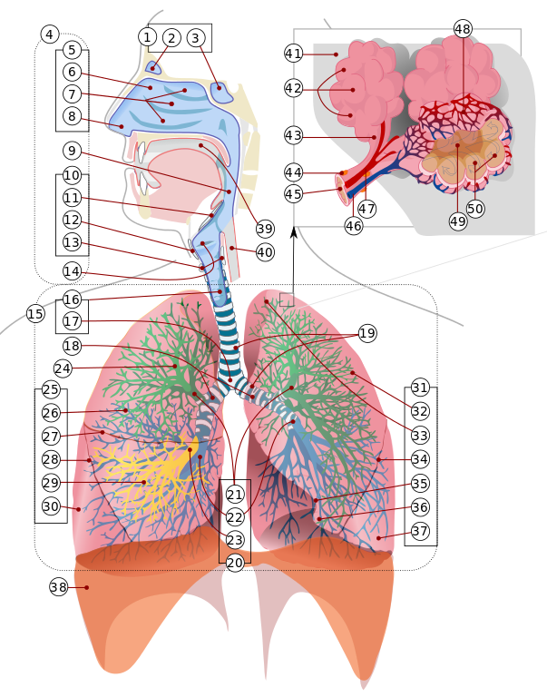
1: Paranasal sinuses (2: Frontal. 3: Sphenoid). 4: Upper respiratory tract: 5: Nose (6: Nasal cavity. 7: Nasal conchae. 8: Nasal vestibule). 9: Pharynx. 10: Larynx (11: Epiglottis. 12: Thyroid cartilage. 13: Cricoid cartilage). 14: Vocal folds. 15: Lower respiratory tract: 16: Trachea (17: Carina). Bronchi (18: Main bronchi. 19: Tracheal and bronchi rings. 20: Lobar bronchus (21: Superior. 22: Inferior. 23: Middle). 24: Lingular division bronchi). 25: Right lung (26: Superior lobe 27: Horizontal fissure. 28: Oblique fissure. 29: Middle lobe. 30: Inferior lobe). 31: Left lung (32: Superior lobe. 33: Apex of left lung. 34: Oblique fissure. 35: Cardiac notch. 36: Lingula of lung. 37: Inferior lobe). 38: Diaphragm. 39: Oral cavity. 40: Esophagus. Respiratory lobule: 41: Connective tissue. 42: Alveolar sacs. 43: Alveolar duct. 44: Mucous gland. 45: Mucosal lining. 46: Pulmonary artery. 47: Pulmonary vein. 48: Capilllary beds. 49: Atrium. 50: Alveoli. |
| Annotations | This image is annotated: View the annotations at Commons |
Faylın tarixçəsi
Faylın əvvəlki versiyasını görmək üçün gün/tarix bölməsindəki tarixlərə klikləyin.
| Tarix/Vaxt | Kiçik şəkil | Ölçülər | İstifadəçi | Şərh | |
|---|---|---|---|---|---|
| indiki | 19:33, 14 fevral 2016 | | 718 × 914 (507 KB) | Jmarchn | Fixed error 43 arrow |
| 00:35, 13 fevral 2016 |  | 718 × 914 (507 KB) | Jmarchn | Renumbered any bronchi | |
| 23:45, 12 fevral 2016 |  | 718 × 914 (507 KB) | Jmarchn | Grouping numbers | |
| 23:30, 11 fevral 2016 |  | 718 × 914 (432 KB) | Jmarchn | A lot of changes in upper respiratory tract and head | |
| 19:27, 13 dekabr 2007 |  | 800 × 900 (330 KB) | LadyofHats | {{Information |Description=numbered version of Image:Respiratory system complete.svg |Source=self-made |Date=dec 2007 |Author= LadyofHats |Permission=Public domain |other_versions=<gallery> Image:Respiratory system complete.svg|en |
Fayl keçidləri
Bu faylı istifadə edən səhifə yoxdur.
Faylın qlobal istifadəsi
Bu fayl aşağıdakı vikilərdə istifadə olunur:
- bg.wikipedia.org layihəsində istifadəsi
- el.wikipedia.org layihəsində istifadəsi
- eu.wikipedia.org layihəsində istifadəsi
- ml.wikipedia.org layihəsində istifadəsi
- ro.wikipedia.org layihəsində istifadəsi
- uz.wikipedia.org layihəsində istifadəsi